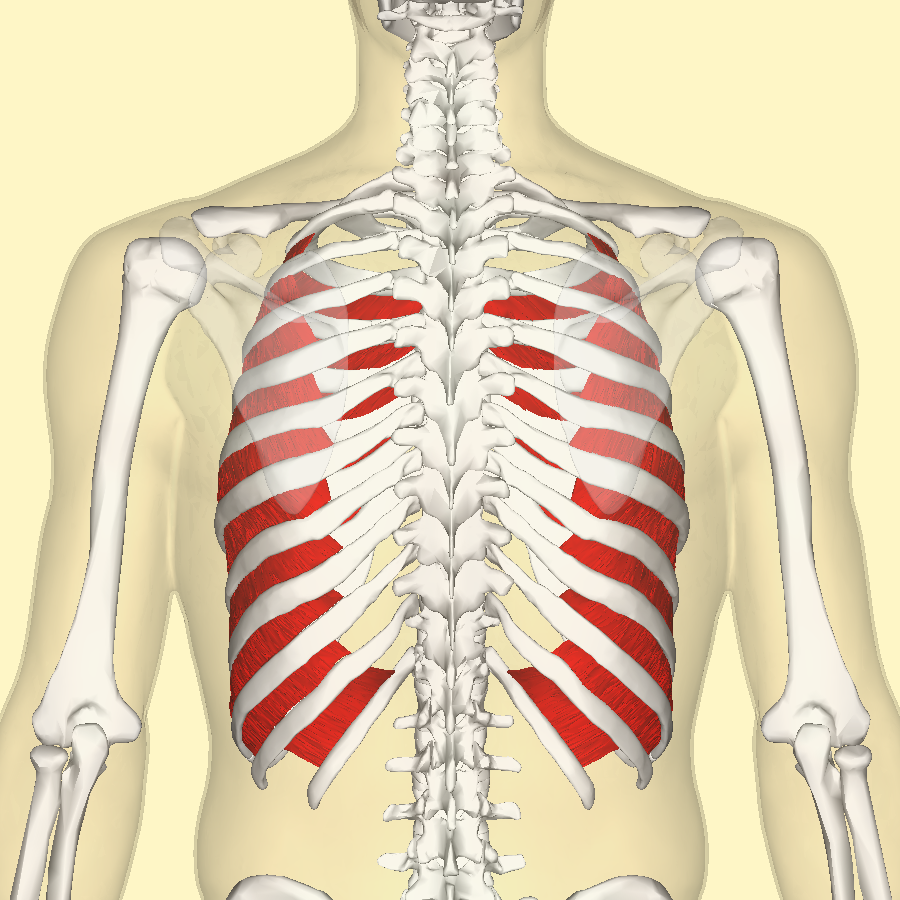
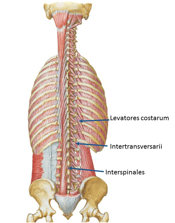
The levatores costarum muscles are a series of epaxial muscles in the thoracic region that receive their nerve supply from the dorsal rami of the spinal nerves. Developmentally, they are modified lateral parts of the intertransversarii muscles that have migrated laterally onto the ribs. Describe the location of the interspinales, intertransversarii, and levatores costarum muscles 5. Describe the origin, insertion, action and nerve supply of the suboccipital muscles 6. Name the boundaries and contents of the suboccipital triangle 7. Describe the origin, course and innervation of the greater occipital nerve. Short levatores costarum muscles and the superior oblique capitis muscle, the latter is also often listed under short neck muscles. Long and short levatores costarum. Image: Levatores costarum. By Uwe Gille, License: Public domain. The 12 long and short levatores costarum muscles originate in the transverse.
- Levatores costarum; longus capitis; longus colli; lumbricals of foot (4) lumbricals of hand; masseter; medial pterygoid; medial rectus; mentalis; m. Uvulae; mylohyoid; nasalis; oblique arytenoid; obliquus capitis inferior; obliquus capitis superior; obturator externus; obturator internus (A) obturator internus (B) omohyoid; opponens digiti.
- Intermediate layer: superior and inferior serratus posterior and the levatores costarum Deep layer: erector spinae, transversospinalis, interspinalis, and the intertransversarii The superficial and intermediate layers receive their nervous supply from peripheral nerves, which are not encountered through the posterior approach ( FIG 2 ).
Levatores Costarum Function
- Superficial landmarks enable gross determination of the anatomic level. Proximally, C7 and T1 are the largest spinous processes and may serve as palpable anatomic landmarks. Distally, the intercrestal line approximates the L4-L5 interspace.
- There are three layers to the posterior musculature of the spine (FIG 1; Table 1):
- Superficial layer: trapezius, latissimus dorsi, rhomboid major and minor, and the levator scapulae
- Intermediate layer: superior and inferior serratus posterior and the levatores costarum
- Deep layer: erector spinae, transversospinalis, interspinalis, and the intertransversarii
- The superficial and intermediate layers receive their nervous supply from peripheral nerves, which are not encountered through the posterior approach (FIG 2). The deep layer receives its nervous supply segmentally from the posterior dorsal rami. There is a large amount of redundancy in the innervation of the deep layer.
- The midline approach is a true internervous plane. Nerve injury occurs only with excessive lateral dissection.
- The vascular supply to the deep layer is from segmental branches of the aorta. These vessels enter the operative field at the level of the intertransverse ligament and can be a source of significant bleeding.
- The facet joint capsules have a shiny white appearance and the individual fibers can be seen inserting onto the lateral edge of the laminar trough. Care should be taken to avoid violating the capsular fibers, unless that segment is being fused.
- The ligamentum flavum has a yellow appearance with the fibers running in a cephalocaudal direction. The cephalad end of the ligament has a broad insertion from the base of the spinous process to between 50% and 70% of the anterior surface of the lamina. The caudal end of the ligament inserts from the superior edge of the lamina to between 2 and 6 mm of the anterior surface of the lamina.4
- Particularly at the L5-S1 level, the interspace may be widened or the posterior bony anatomy only partly formed. Care should be exercised when exposing this level as inadvertent plunging into the canal may occur.
- Laterally, the intertransverse membrane overlies the iliopsoas and protects the neural structures that lie beneath.
- In children, the spinous process apophysis has not fused. During dissection, the apophysis is split down to the bone and then elevated with the paraspinal musculature.
FIG 1 • The superficial, intermediate, and deep musculature of the back. |
| |||||||||||||||||||||||||||||||||||||||||||||||||||||||||||||||||||||||||||||||||||||||||||||||||||||||||
You may also need
Muscles of the back can be divided into superficial, intermediate, and deep group.
Since the all the back muscles originate in embryo (fetus) form by locations other than the back, muscles in the superficial, as well as, intermediate groups, are extrinsic muscles. Anterior rami of spinal nerve innervate them.
Muscles associated and involved in movements of the upper limb belong to the superficial group. The intermediate group might perform a respiratory action and comprises of muscles connected to the ribs.
The deep group of muscles is called intrinsic muscles because they develop in the back. They are direct association with actions of the vertebral column and head and are supplied by posterior rami of spinal nerves.
Superficial Group of Back Muscles
Superficial Layer of Back Muscle
Superficial muscles of the back are located directly deep towards the skin along with superficial fascia. They are occasionally called the appendicular group as these muscles are mainly associated with activities of the appendicular skeleton. The superior part of the appendicular skeleton that includes clavicle, scapula, and humerus, is attached to the axial skeleton that consists of skull, ribs, and vertebral column by these muscles.
Trapezius
Origin: Spinous prominences of CVII to TXII, Superior nuchal line, external occipital protuberance, ligamentum nuchae.
Insertion: Lateral one third of clavicle, acromion, and spine of scapula.
Innervation: Motor stimulation is due to accessory nerve [XI], and sensory stimulation is due to sensory nerve endings C3 and C4.
Functions: During abduction of humerus above horizontal it assists in rotating the scapula; upper fibers help in elevation, middle fibers help in adduction, and lower fibers help in depression scapula.
Latissimus Dorsi
Origin: Spinous processes of TVII to LV and sacrum, iliac crest, ribs X to XII.
Insertion: Floor of intertubercular sulcus of humerus.
Innervation: Thoracodorsal nerve (C6 to C8).
Functions: Extends, adducts, and medially rotates humerus.
Levator Scapulae
Origin: Transverse processes of CI to CIV.
Insertion: Upper portion medial border of scapula.
Innervation: C3 toC4 and dorsal scapular nerve (C4, C5).
Functions: Elevates scapula.
Rhomboid Major
Origin: Spinous processes of TII to TV.
Insertion: Medial border of scapula between spine and inferior angle.
Innervation: Dorsal scapular nerve (C4, C5).
Functions: Retracts (adducts) and elevates scapula.
Rhomboid Minor
Origin: Lower portion of ligamentum nuchae, spinous processes of CVII and TI.
Insertion: Medial border of scapula at spine of scapula.
Innervation: Dorsal scapular nerve (C4, C5).
Functions: Retracts (adducts) and elevates scapula.
Intermediate Group of Back Muscles
Both Serratus posterior superior and Serratus posterior inferior move obliquely outwards via the vertebral column to connect to the ribs.
Serratus posterior superior is located deep to the rhomboid muscles, while serratus posterior inferior is located deep to the latissimus dorsi. Both serratus posterior muscles are attached to the vertebral column and associated structures medially and either descends like the fibers of the serratus posterior superior or else ascend the fibers of the serratus posterior inferior to connect to the ribs.
These two muscles elevate and depress the ribs. Segmental branches of anterior rami of intercostal nervesinnervate the serratus posterior muscles. Intercostal arteries give vascular supply to them.
Serratus Posterior Superior
Origin: Lower portion of ligamentum nuchae, spinous processes of CVII to TIII, and supraspinous ligaments.
Insertion: Upper border of ribs II to V just lateral to their angles.
Innervation: Anterior rami of upper thoracic nerves (T2 to T5).
Function: Elevates ribs II to V.
Serratus Posterior Inferior
:watermark(/images/watermark_only.png,0,0,0):watermark(/images/logo_url.png,-10,-10,0):format(jpeg)/images/anatomy_term/musculi-intertransversarii-anteriores-et-posteriores-cervicis/P1YA6nmsfp4OwVn5D6ClNA_Musculi_intertransversarii_anteriores_et_posteriores_cervicis_1.png)
Intermediate Layer of Back Muscles
Origin: Spinous processes of TXI to LIII and supraspinous ligaments.
Insertion: Lower border of ribs IX to XII just lateral to their angles.
Innervation: Anterior rami of lower thoracic nerves (T9 to T12).
Function: Depresses ribs IX to XII and may prevent lower ribs from being elevated when the diaphragm contracts.
Deep group of back muscles
The deep or intrinsic muscles of the back extend from the pelvis to the skull and are innervated by segmental branches of the posterior rami of spinal nerves. They consist of:
- The extensors and rotators of the head and neck: the splenius capitis and cervicis (spinotransversales muscles).
- The extensors and rotators of the vertebral column: the erector spinae and transversospinales. and
- The short segmental muscles: the interspinales and intertransversarii.
The vascular supply to this deep group of muscles is through branches arteries like:
- Vertebral
- Deep cervical
- Occipital
- Transverse cervical
- Posterior intercostal
- Subcostal
- Lumbar
- Lateral sacral arteries
Spinotransversales Muscles
The two spinotransversales muscles go upward and laterally via the spinous processes and ligamentum nuchae:
- The splenius capitis is a broad muscle attached to the occipital bone and mastoid process of the temporal bone.
- The splenius cervicis is a narrow muscle attached to the transverse processes of the upper cervical vertebrae.
Independently, each muscle turns the head to one side that is the same side as the contracting muscle and collectively, the spinotransversales muscles move the head backward, elongating the neck.
Splenius Capitis
Origin: Lower half of ligamentum nuchae, spinous processes of CVII to TIV.
Insertion: Mastoid process, skull below lateral one third of superior nuchal line.
Innervation: Posterior rami of middle cervical nerves.
Function: Together, they draw head backward, extending the neck and individually, each one draws and rotates head to one side i.e. turns face to same side.
Splenius Cervicis
Origin: Spinous processes of TIII to TVI.
Insertion: Transverse processes of CI to CIII.
Innervation: Posterior rami of lower cervical nerves.
Function: Together they extend neck, and individually they draw and rotate head to one side i.e. same side.
Erector Spinae Muscles
Among intrinsic back muscles the erector spinae remains the largest group. The muscles are located medially amid the spinous processes, posterolaterally towards the vertebral column and laterally towards the angles of the ribs. Inside the thoracic and lumbar regions thoracolumbar fascia as well as the serratus posterior inferior, the rhomboid and splenius muscles surround them.
Muscles of the Back: Erector Spinae Muscles
Iliocostalis Lumborum
It originates via sacrum, spinous processes of lumbar and lower two thoracic vertebrae and their supraspinous ligaments, and the iliac crest; and Insert inside angles of the lower six or seven ribs.
Iliocostalis Thoracis
It originates in Angles of the lower six ribs and inserts in angles of the upper six ribs and the transverse process of CVII.
Iliocostalis Cervicis
It originates in angles of ribs III to VI, while Inserts in Transverse processes of CIV to CVI.
Longissimus Thoracis
It starts in blends with iliocostalis in lumbar region and is attached to transverse processes of lumbar vertebrae. While it Inserts in Transverse processes of all thoracic vertebrae, towards the tubercles of the lower nine or ten ribs laterally.
Longissimus Cervicis
It originates in transverse processes of upper four or five thoracic vertebrae and it inserts in transverse processes of CII to CVI.
Longissimus Capitis
It originates in Transverse processes of upper four or five thoracic vertebrae and articular processes of lower three or four cervical vertebrae and inserts in posterior margin of the mastoid process.
Spinalis Thoracis
It originates in Spinous processes of TX or TXI to LII and inserts in spinous processes of TI to TVIII (varies).
Spinalis Cervicis
It originates in lower part of ligamentum nuchae and spinous process of CVII (sometimes TI to TII) and inserts in spinous process of CII (axis).
Spinalis Capitis
It originates while blending with semispinalis capitis and it inserts in semispinalis capitis.
The transversospinales muscles go from transverse processes to spinous processes occupying the groove between these two vertebral projections obliquely upward and medially. They are made up of of three major subgroups the semispinalis, multifidus, as well as rotatores muscles and are situated deep towards the erector spinae.
- The semispinalis muscles are the most superficial collection of muscle fibers in the transversospinales group.
- Deep to the semispinalis is the second group of muscles, the multifidus.
- The small rotatores muscles are the deepest of the transversospinales group.
Semispinalis Thoracis
It originates inside transverse processes of TVI to TX and inserts in spinous processes of upper four thoracic and lower two cervical vertebrae.

Semispinalis Cervicis
It originates in transverse processes of upper five or six thoracic vertebrae and inserts in spinous processes of CII (axis) to CV.
Semispinalis Capitis
It originates in transverse processes of TI to TVI (or TVII) and CVII and articular processes of CIV to CVI, and inserts in Medial area between the superior and inferior nuchal lines of occipital bone.
Multifidus
It originates through Sacrum, origin of erector spinae, posterior superior iliac spine, mammillary processes of lumbar vertebrae, transverse processes of thoracic vertebrae, and articular processes of lower four cervical vertebrae while it inserts in the base of spinous processes of all vertebrae from LV to CII (axis).
Rotatores Lumborum
It starts in transverse processes of lumbar vertebrae and inserts in spinous processes of lumbar vertebrae.
Rotatores Thoracis
It originates in transverse processes of thoracic vertebrae and Inserts spinous processes of thoracic vertebrae.
Rotatores Cervicis
It originates in articular processes of cervical vertebrae and inserts in spinous processes of cervical vertebrae.
Segmental Muscles

The two groups of segmental muscles are innervated via posterior rami of spinal nerves and are located deeply in the back. The first group of segmental muscles that arise via the transverse processes of vertebrae CVII and TI to TXI is the levatores costarum muscles. They insert within the rib under the vertebra of origin in the area of the tubercle and have an oblique lateral and downward course. Their contraction elevates the ribs. The second group of segmental muscles are called the interspinales and the intertransversarii, which pass between neighboring spinous processes is the real segmental muscles of the back, which pass between adjacent transverse processes. They stabilize surrounding vertebrae to enable more efficient activity of the large muscle groups in actions of the vertebral column.
Muscles of the Back – Segmental Muscles
Levatores Costarum
Interspinales And Intertransversarii
It originates short paired muscles arising from transverse processes of CVII to TXI and inserts the rib below vertebra of origin near tubercle. Its main function is elevation of ribs through contraction.
Interspinales
It originates in short paired muscles attached to the spinous processes of contiguous vertebrae, one on each side of the interspinous ligament and its main function as postural muscle is that it stabilizes adjoining vertebrae during movements of vertebral column.
Intertransversarii
It originates in small muscles between the transverse processes of contiguous vertebrae. Its primary function as postural muscle is stabilizing adjoining vertebrae during movements of vertebral column.